Vatech Pax I OneShot
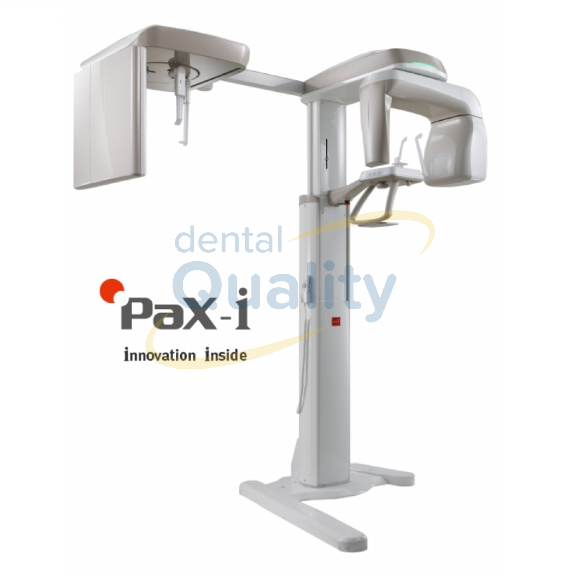
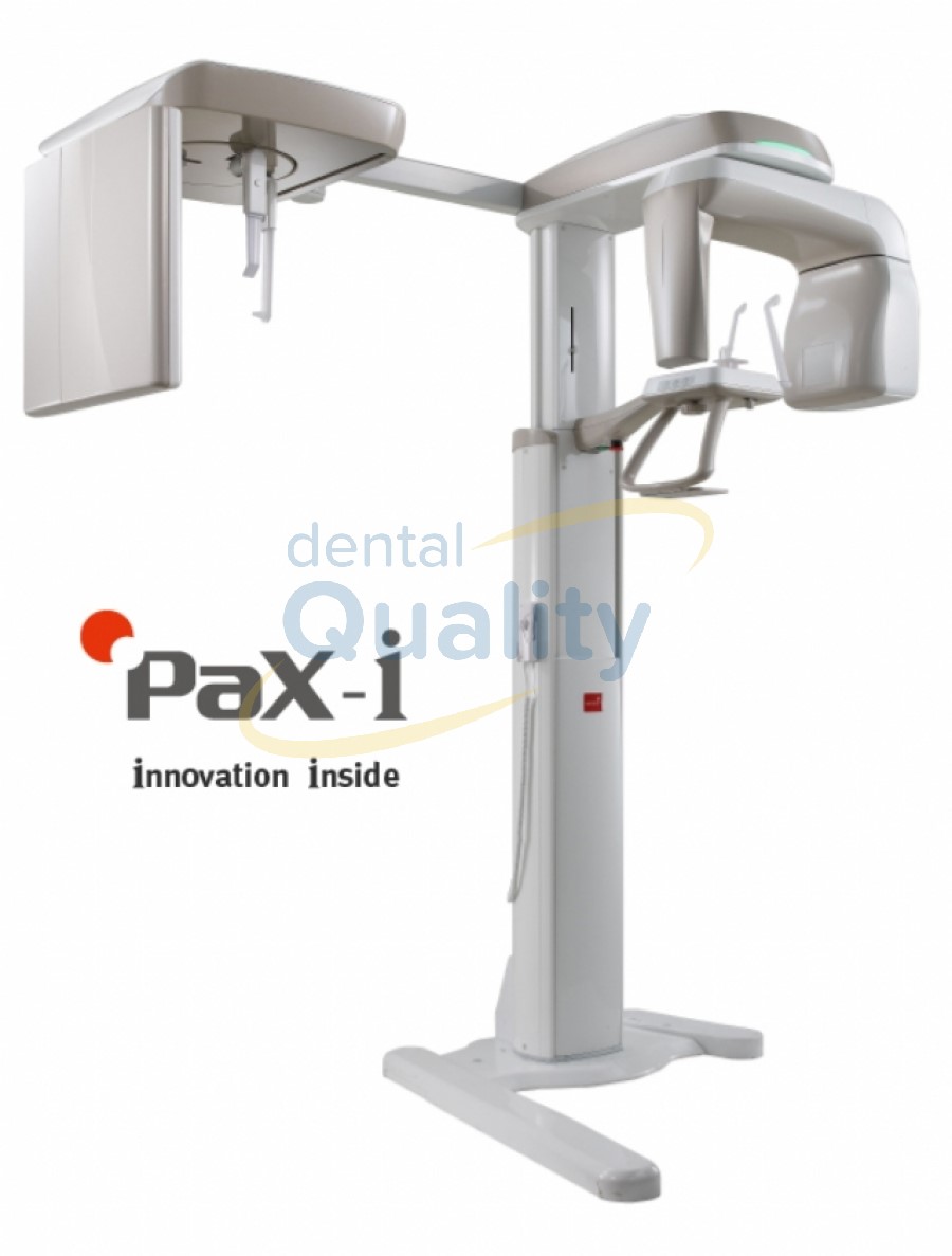
Optimal image for accurate diagnosis
Specialized sensors for Pano & Ceph
Streamlined workflow and prolonged lifespan of sensors
Raise design value of your clinic
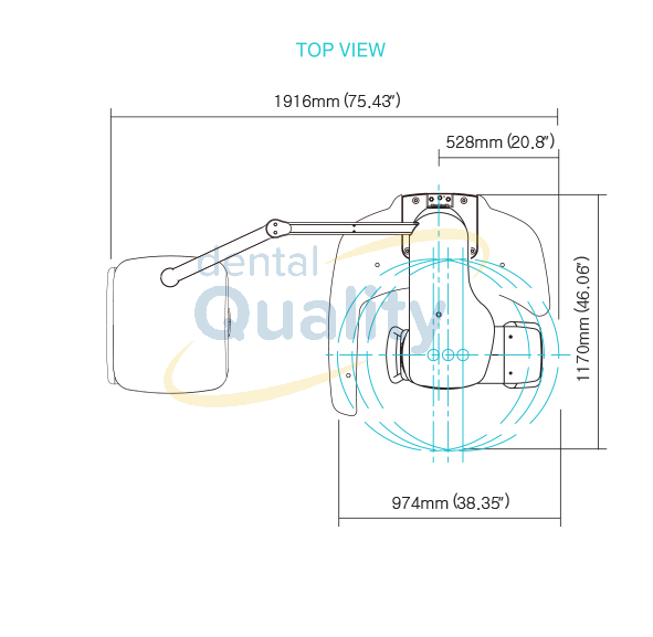
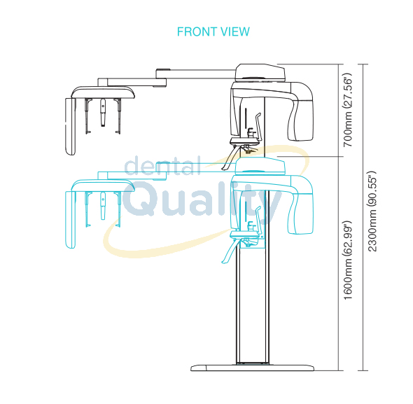
Save installation space in your clinic
Panoramic
Standard / Right / Front / Left
Orthogonal Standard / Right / Front / Left Bitewing Standard / Right / Front / Left
TMJ LAT Open / Close
TMJ PA Open / Close
Sinus LAT / PA
Cephalometric
PaX-i Provides optimal images with an exclusively designed sensor for cephalometric diagnosis.
As it offers two image sizes, LAT and Full LAT, you can choose one of them based on the purposes of your diagnostic needs.
LATERAL
Provide specialized high quality images to suit orthodontics occipital and maxillofacial surgeries. diagnosis. (optional)
FULL LATERAL
Full lateral gives 30% larger images and the area of the patient for comprehensive diagnosis. (optional)
Three different ceph image sizes reduce unnecessary X-ray dosage and scans the ideal area of cranial anatomy for your diagnosis and treatment planning.
Function Pano + Ceph
Scan Time Pano : 13.5 sec (HD) / Normal 10.1 sec
Ceph : Scan 12.94 sec / One-shot 0.9 sec
Focal Spot 0.5 mm
Tube Voltage / Current Pano : 50~90 Kvp / 4-10 mA
Ceph FOV Size SC : 21x23cm (8.3x9.1") [LAT, PA, SMV, Water View, Carpus]
27x23cm (10.6x9.1") [Full LAT]
OS : 23x25cm (9x10") [LAT, PA, SMV, Water View, Carpus]
OP : 30x25cm (12x10") [LAT, PA, SMV, Water View, Carpus]
Gray Scale 14 bit
Patient Position Standing / Wheel-chair accessible
Specialized sensors for Pano & Ceph
Streamlined workflow and prolonged lifespan of sensors
Raise design value of your clinic
Save installation space in your clinic
Panoramic
Standard / Right / Front / Left
Orthogonal Standard / Right / Front / Left Bitewing Standard / Right / Front / Left
TMJ LAT Open / Close
TMJ PA Open / Close
Sinus LAT / PA
Cephalometric
PaX-i Provides optimal images with an exclusively designed sensor for cephalometric diagnosis.
As it offers two image sizes, LAT and Full LAT, you can choose one of them based on the purposes of your diagnostic needs.
LATERAL
Provide specialized high quality images to suit orthodontics occipital and maxillofacial surgeries. diagnosis. (optional)
FULL LATERAL
Full lateral gives 30% larger images and the area of the patient for comprehensive diagnosis. (optional)
Three different ceph image sizes reduce unnecessary X-ray dosage and scans the ideal area of cranial anatomy for your diagnosis and treatment planning.
Function Pano + Ceph
Scan Time Pano : 13.5 sec (HD) / Normal 10.1 sec
Ceph : Scan 12.94 sec / One-shot 0.9 sec
Focal Spot 0.5 mm
Tube Voltage / Current Pano : 50~90 Kvp / 4-10 mA
Ceph FOV Size SC : 21x23cm (8.3x9.1") [LAT, PA, SMV, Water View, Carpus]
27x23cm (10.6x9.1") [Full LAT]
OS : 23x25cm (9x10") [LAT, PA, SMV, Water View, Carpus]
OP : 30x25cm (12x10") [LAT, PA, SMV, Water View, Carpus]
Gray Scale 14 bit
Patient Position Standing / Wheel-chair accessible
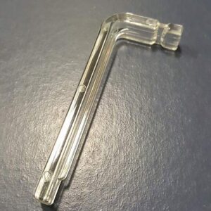
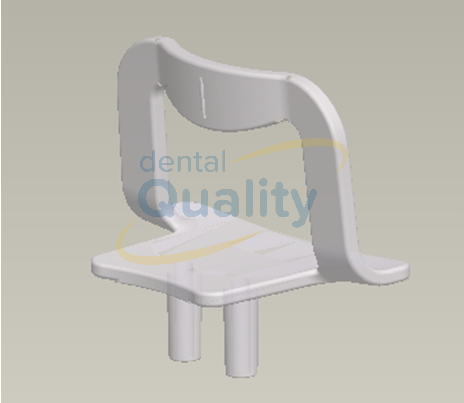
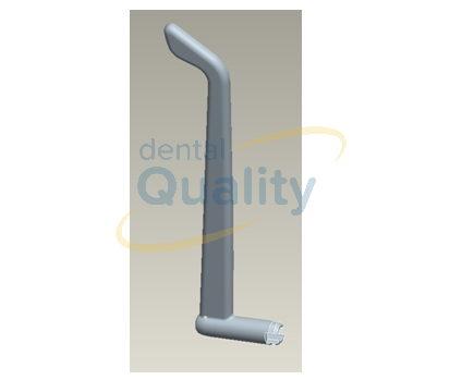
Support temporal gauche Pax I SP/SC/OP
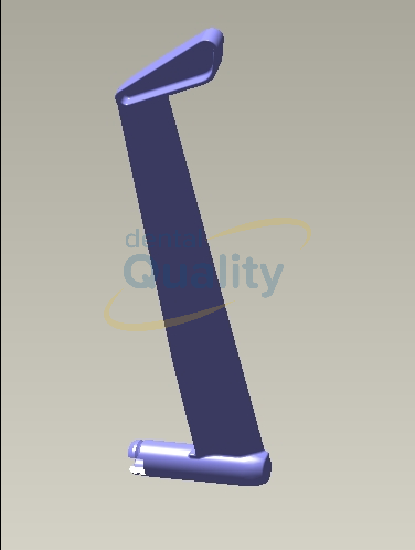
Support temporal droit Pax I SP/SC/OP
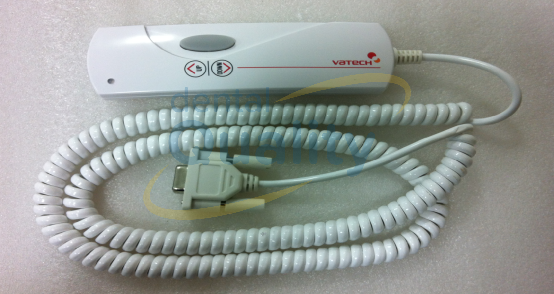
Commande monter descente Pax I SP/SC/OP
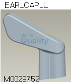
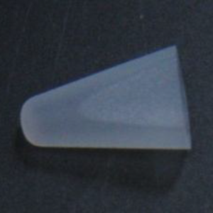
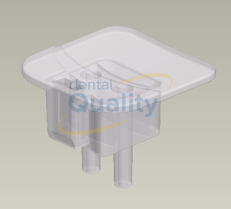
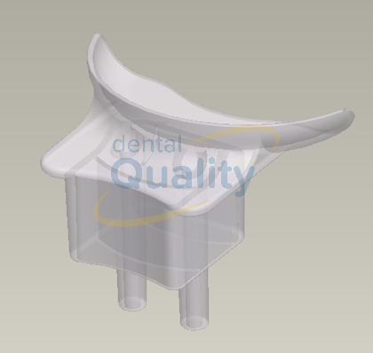
SILICON-CAP EAR CHIN-L/SILICON CLEAR/M0029752
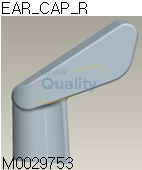
SILICON-CAP EAR CHIN-R/SILICON CLEAR/M0029753